Precise developmental gene expression arises from globally stochastic transcriptional activity
Shawn C. Little, Mikhail Tikhonov and Thomas Gregor, Cell 154, 789–800 (2013).
Abstract
Early embryonic patterning events are strikingly precise, a fact that appears incompatible with the stochastic gene expression observed across phyla. Using single molecule mRNA quantification in Drosophila embryos, we determine the magnitude of fluctuations in the expression of four critical patterning genes. The accumulation of mRNAs is identical across genes and fluctuates by only ~8% between neighboring nuclei, generating precise protein distributions. In contrast, transcribing loci exhibit an intrinsic noise of ~45% independent of specific promoter-enhancer architecture or fluctuating inputs. Precise transcript distribution in the syncytium is recovered via straightforward spatiotemporal averaging—that is, accumulation and diffusion of transcripts during nuclear cycles—without regulatory feedback. Common expression characteristics shared between genes suggest that fluctuations in mRNA production are context-independent and are a fundamental property of transcription. The findings shed light on how the apparent paradox between stochastic transcription and developmental precision is resolved.
Synopsis
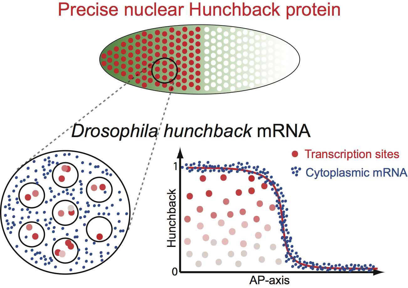 Single-molecule quantification in Drosophila embryos reveals that a paradox between extremely precise embryonic patterning and stochastic transcription is resolved by simple physical averaging mechanisms.
Highlights
- Absolute quantification of nascent and mature mRNA in Drosophila embryos
- Nascent transcription is noisy, whereas cytoplasmic mRNA levels are precise
- All early expressed genes exhibit the same degree of transcriptional variability
- Spatiotemporal averaging across multiple genomic loci generates precision
View a high-resolution image stack of wild-type embryo labeled hunchback mRNA
Video still 1:
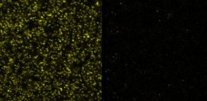 Anterior (left) and posterior (right) 25×25 μm² region of wild-type embryo (staged during interphase of the 13th nuclear cycle) stained with hb smFISH probes. Imaging depth is just below the nuclear layer, approximately 10 μm below the surface.
Video still 2:
 Anterior (left) and posterior (right) 25×25 μm² region of wild-type embryo (staged during interphase of the 13th nuclear cycle) stained with hb smFISH probes. Imaging depth is in the nuclear layer, approximately 7 μm below the surface.
In the anterior image the dark outlines of nuclei devoid of cytoplasmic transcripts can be seen. Bright spots inside nuclei correspond to sites of nascent transcription (many (i.e. 10-100) unfinished mRNA molecules amplify signal here). Single cytoplasmic transcripts can be seen in inter-nuclear spaces.